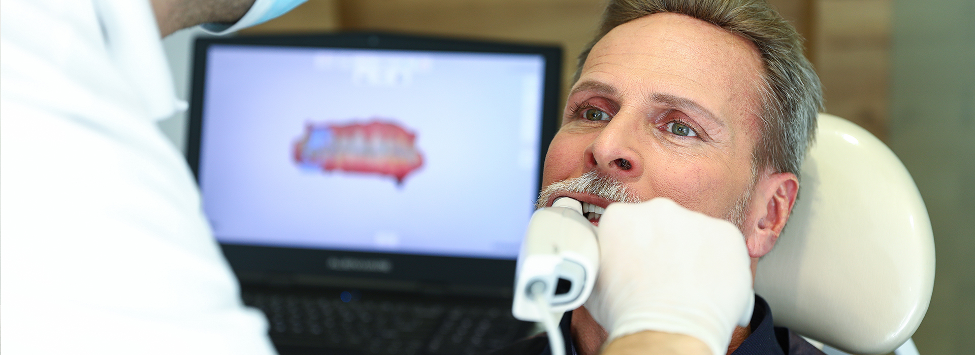
Dental X-ray in New York

A comprehensive examination makes it possible to make the correct diagnosis and choose the required treatment method. Even a professional, experienced dentist sees only 50% of the dental tissue on visual inspection. The remaining half can be seen using an X-ray of the jaw or a panoramic picture of the teeth. This research method allows you to get a detailed picture of the bones of the jaw and teeth for further diagnosis and treatment of diseases. The general condition of the jaw and nearby tissues, the location of the roots of the dental system, and its other fragments – all this is shown by radiography.

Dentists of iSmile Dental have prepared a list of medical procedures before which a good doctor will necessarily prescribe an X-ray of the teeth.

Procedures for which X-ray is required.
  1. Upcoming implantation. With the help of X-ray, it is possible to assess bone tissue, that is, the width and height of the lower jaw, the distance to the mandibular nerve, and the maxillary sinuses on the upper jaw.

  2.    Prosthetics. It is necessary to develop the proper step-by-step treatment.

  3.   Installation of braces. Drawing up a correct plan for step-by-step correction of the bite is necessary.

  4.   Complex tooth extraction (for example, wisdom teeth). It is necessary to draw up an optimal treatment plan.

  5.   Detection of the stage of periodontal disease. X-ray teeth are prescribed if necessary to determine the depth of periodontal pockets.

  6.   With inflammation of bone tissue. This procedure is recommended if it is necessary to detect the focus of inflammation.

  7.   Control of tooth growth in young children. It helps monitor the bite's development in time to detect and correct the problem.

  8.   In some cases, the X-ray procedure may be prescribed by an ENT. This is usually necessary to establish whether there is a connection between the inflammation of the tooth and a severe form of sinusitis.
Read More Read Less

Advantages and disadvantages of X-ray diagnostics.

X-ray examination has many advantages and is practically devoid of disadvantages. The main benefits are - the speed and simplicity of the procedure; the almost complete absence of contraindications; the high information content of the research results; lack of any uncomfortable sensations in the patient; there is no need for complex preparation for the procedure. Separately, the result of the study can be obtained without waiting.

Of the method's shortcomings, it can only be noted that the studied area is irradiated with X-ray radiation during the examination. And even though the intensity of such radiation is minimal and can not harm a person, the number of X-ray examinations per year is limited.

There are practically no contraindications for the radiography of teeth and jaws. The procedure is safe and absolutely painless. Modern X-ray machines, as before, irradiate with radioactive radiation, but the irradiation values are so small that they do not affect human health in any way. The only exception may be pregnant women, who are advised to refrain from carrying out this procedure until the end of the second trimester.

X-ray of teeth gives a 100% accurate result. iSmile Dental dentists provide the patient with the result in any convenient format: on paper, X-ray film, or CD-ROM.

Read More Read Less

faq

If the doctor needs to get precise information about the entire oral cavity and not just individual teeth, then it is best to make an orthopantomogram. This is the same X-ray, but all bone formations are visible from the lower jaw to the maxillary sinuses.

An orthopantomogram is a 2D picture of the teeth of the upper and lower jaw and the temporomandibular joint. A panoramic image allows you to see and assess the patient’s teeth’ condition.

Yes, you can make an Rg-gram of a tooth if you know its number without a doctor’s referral. The cost depends on the study type and carrier (whether it is performed on Rg-film or a computer study with a printout).

Modern dental digital tomographs are easy to use and provide high-quality information. The radiation dose with computed tomography is less than with a panoramic image taken on a film orthopantomograph and is approximately 45 microsieverts.

The information content (validity) of the orthopantomogram is a maximum of 1 year.

Dentists treat teeth without X-rays only in the case of apparent caries in the absence of hidden causes. An X-ray will help the doctor determine the cause of the toothache and the place and form of the inflammatory process.

The procedure takes no more than 1-2 minutes, but the sequence of actions depends on the type of X-ray. With intraoral radiography, a lead apron covering the chest is preliminarily put on the patient, brought to the device, a piece of film or a digital sensor is applied to the X-ray area, and a picture is taken.

The purpose of radiography in dentistry is to make an accurate diagnosis or determine the treatment plan. Most often, manipulation is prescribed in such cases – suspicion of granulomas, cysts near the tooth root, deep caries, and other dental diseases.

If we talk about how often you can do an X-ray of the teeth of an adult without harm to health, then the optimal response will be 3-5 times a month (if required). In general, the dose of X-rays of the teeth should not exceed 150 mSv per year.

An experienced dentist always knows what caries looks like on an X-ray and determines it without problems. Carious cavities X-rays show in the form of darkening. In the initial stages of caries, they are not pronounced and are localized within the dentin.

Request Booking or Consultation Online